
Figure 5.
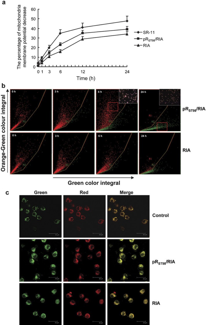
Measurement of mitochondrial membrane potential of J774A.1 cells. (a) The percentage of mitochondria membrane potential (Δψm) decrease in J774A.1 cells. At 1 h post-infection, S. typhimurium strain pRST98/RIA resulted in a higher number of J774A.1 with decreased mitochondrial Δψm than RIA, and a time-dependent decrease in mitochondrial Δψm was observed. (b, c) Fluorescent JC-1 reaction examination of J774A.1 cells undergoing mitochondria Δψm decrease detected with a laser scanning cytometer (×200) and laser scanning confocal microscope, respectively. The corner panels are higher magnifications of the boxed areas. S. typhimurium, Salmonella enterica serovar Typhimurium.