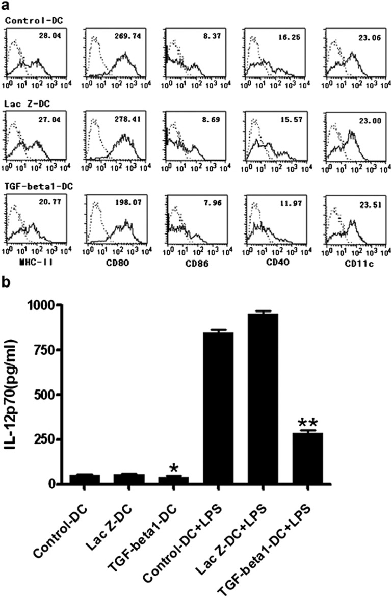
Figure 2.
Characteristics of TGF-β1-DCs. (a) Flow cytometric analysis of murine DCs. After infection of adenovirus for 48 h, each group of DCs was stained with FITC-coupled antibodies specific for MHC-II, CD11c, CD80, CD86, or the corresponding isotype controls. (b) Levels of IL-12p70 in the culture supernatant of TGF-β1 gene-modified immature DCs. Six-day-old DCs were infected with Ad-TGF-β1 or Ad-LacZ at an MOI of 50 for 24 h in serum-free medium. After washing five times, the cells were stimulated with LPS (100 ng/ml) for 20 h. Levels of IL-12p70 in the culture supernatants were determined by ELISA. *P<0.05, **P<0.01; data are representative of three independent experiments (n=3). Ad, adenoviral; DC, dendritic cell; LPS, lipopolysaccharide; MOI, multiplicity of infection; TGF-β1, transforming growth factor-β1.

