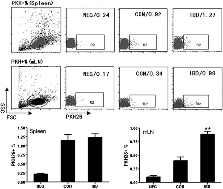
Figure 6.
Lymphocytes from mLNs of IBD mice recruit more TGF-β1-DCs than those from normal mice. DCs were labeled with PKH26 and injected into normal mice (CON) and IBD model mice (IBD). NEG was a group of normal mice injected with unlabeled DCs as negative control. The mice were killed 24 h after injection, and the splenocytes and lymphocytes from mLNs were collected and subjected to FACS analysis. **P<0.01; data are representative of three independent experiments (n=3). DC, dendritic cell; IBD, inflammatory bowel disease; mLN, mesentery lymph node; FACS, fluorescence-activated cell sorter.

