Figure 6.

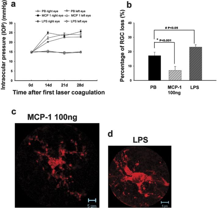
The effects of MCP-1/CCL2 and LPS administered intravitreally in the OH eyes at 4 weeks after the first laser photocoagulation. IOP of the right eyes in different groups showed no significant difference (a). There were significant differences in the percentage of RGC loss when comparing the 100 ng MCP-1/CCL2 and LPS groups with the PB control group (b). Error bars represent the SEM. At 4 weeks after the first laser photocoagulation, iba-1-positive microglia showed a semiactivated state (c) in the 100 ng MCP-1/CCL2-treated retinas while they showed a fully activated phenotype (d) in the LPS-treated retinas. LPS, lipopolysaccharide; MCP-1, monocyte chemoattractant protein-1; PB, phosphate buffer; RGC, retinal ganglion cell; SEM, standard error of the mean.
