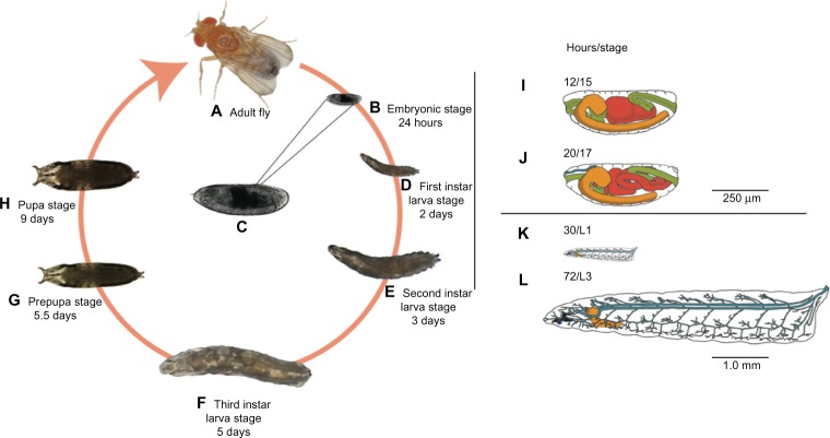
Figure 2.
Drosophila life cycle.
Notes: All stages of the Drosophila life cycle are readily accessible and amenable to manipulation with a variety of basic to high-end tools and techniques. Imaging techniques can be applied in every stage of development (A–H), thanks to the clear cuticle during embryonic (B and C) and larval (D–F) stages. Here we use stage 15 embryos (C), which correspond to roughly 50% of completed embryonic development. Under ideal growing conditions, this stage is reached approximately 12 hours after egg laying and features a developing central nervous system (orange), digestive tract (green and red), and many other systems (not shown) with development underway (I). In stage 15, the midgut has one compartment that divides into two distinct compartments as the embryo progresses to stage 16. We used this feature as an indication of initial survival after nanoparticle delivery and used morphological features characteristic of later developmental stages (J–L) for mortality determinations. For a detailed review of these morphological features, please see Campos-Ortega and Hartenstein.41 Note that time points in the figure correspond to time elapsed from egg laying to the end of a particular developmental stage.

