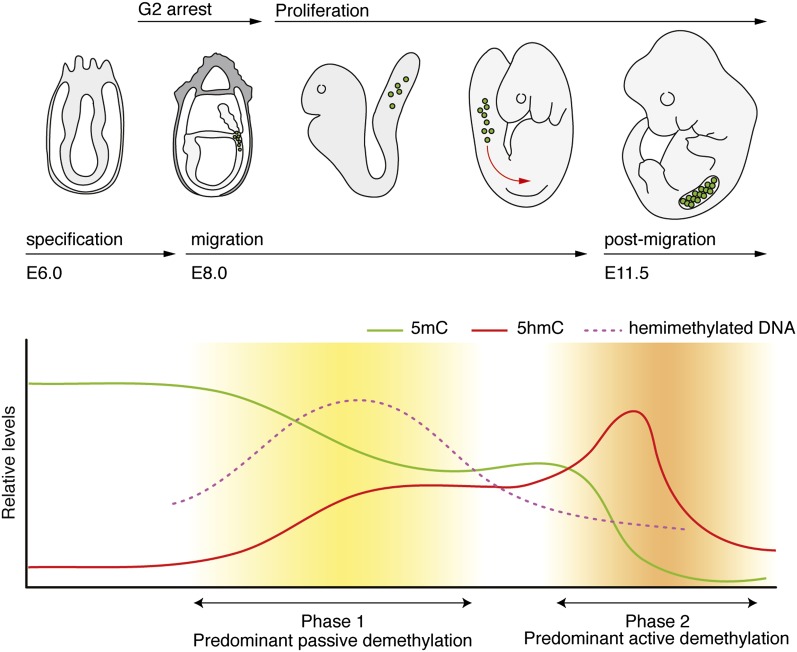
Figure 3.
Biphasic demethylation dynamics in mouse PGCs. PGCs are derived from the embryonic ectoderm of the E6.5 embryo and display high (somatic) 5mC levels (green lines) and low 5hmC levels (red lines). Upon migration, PGCs proliferate, and 5mC levels are passively diluted. Coincidently, hemimethylated DNA strands accumulate transiently and are subsequently lost (purple dashed line). Post-migratory PGCs enter a phase of active DNA demethylation, resulting in an almost complete loss of 5mC and a transient enrichment of 5hmC. At E13.5, both 5mC and 5hmC levels are low.

