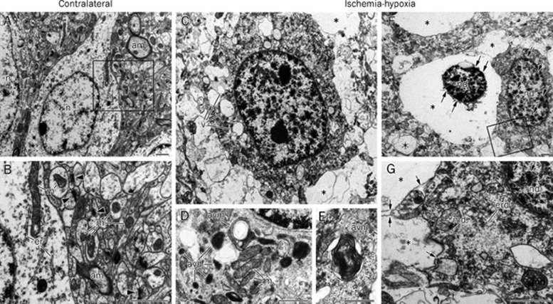
Figure 2.
Ischemia-hypoxia induces vacuolization and lysis of cellular organelles. A and B: Contralateral hemisphere illustrates normal structures of neuronal cell bodies and the neuropil. Notice homogenous nuclear chromatin (n), myelinated axons (am), intact mitochondria (m), cisterns of endoplasmic reticulum (er), and numerous synapses (arrowheads). C–G: Damaged neurons in the cerebral cortex at 6 h after ischemia-hypoxia show multiple vacuoles (v in C) in the cytoplasm and different degrees of cell destruction. Electron micrographs of the cytoplasm in less damaged neurons (D and E; notice intact mitochondria, m) exhibit autophagy-like vacuoles containing electron-dense material (avd) and whorls of membranous material (avm). In more advanced cell destruction, many cells demonstrate the near-complete lysis of organelles (asterisks in C, F, and G), but the plasma membrane of such cells is preserved (arrows in G). Moderately damaged cells (G) display fragmented endoplasmic reticulum membranes (erd), condensed chromatin (double arrows) in damaged nuclei (nd), and swollen mitochondria (md) containing an electron transparent matrix. Condensation of chromatin (double arrows in F and G) in damaged nuclei might be the result of a proapoptotic reaction or represent aborted apoptosis. B and G represent the enlarged framed areas in A and F, respectively. Scale bars: 1 μm (A–C, F, G); 0.5 μm (D, E). Used with permission from Adhami et al whose original article was published in the American Journal of Pathology, Volume 169, Issue 2, August 2006, Pages 566–583 (Copyright © 2006 American Society for Investigative Pathology Published by Elsevier Inc)11.

