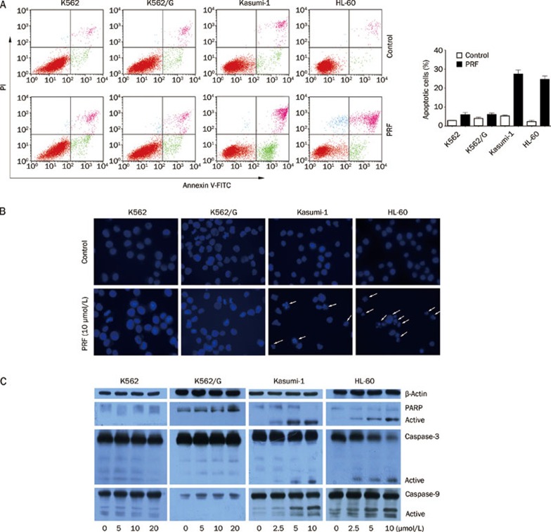
Figure 2.
Apoptosis induced by perifosine in AML cells but not in CML cells. (A) After treatment with perifosine (PRF) at 10 μmol/L for 24 h, leukemia cell lines were harvested and detected by annexin V/propidium (PI)-staining method. The inserted panel shows data from three separate experiments (B) CML and AML cells were stained with Hoechst 33258 from one day after perifosine (10 μmol/L) treatment, and then observed under a fluorescence microscope. Arrows represent the apoptotic nuclei. (C) After incubation of leukemia cell lines for 24 h with the indicated concentrations of perifosine, whole cell extracts were analyzed by Western blot analysis using anti-caspase-3, -9, and PARP antibodies. β-Actin was used as a loading control. The results are representatives of three separate experiments.

