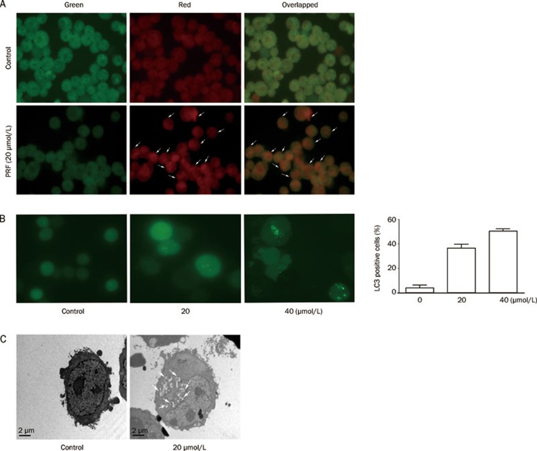
Figure 4.
Induction of autophagy by perifosine(PRF) in K562 cells. (A) K562 cells were treated with or without 20 μmol/L perifosine for 48 h, and then stained with acridine orange (1 μg/mL), and then visualized under green and red filter microscope; note the presence of numerous autophagical vacuoles (white arrows). All digital micrographs were taken at the same exposure setting and images were overlapped. (B) K562 cells that were transfected with GFP-LC3 were incubated for 24 h without or with perifosine at 20 and 40 μmol/L, respectively, and then examined by a fluorescence microscopy. The cells with LC3-positive vesicles were calculated using ImageJ software, and data from three independent experiments were shown (right panel). (C) Representative TEM photomicrographs (×3700) of the K562 cells treated without or with 20 μmol/L perifosine for 48 h. Arrows, autophagic vacuoles. Scale bars, 2 μm.

