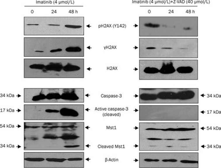
Figure 3.
Caspase-3/Mst1 pathway is involved in imatinib-induced H2AX phosphorylation at Ser139 and Tyr142. K562 cells were treated with imatinib (left panels) and imatinib with Z-VAD (right panel) for the indicated time. The extracted whole proteins were resolved by 10% SDS-PAGE followed by Western blot analysis with antibodies against caspase-3 (full length, 34 kDa), cleaved caspase-3 (17 kDa fragment), Mst1 (full length, 54 kDa), and cleaved Mst1 (34 kDa) (bottom, left panel and right panel). The extracted histones were analyzed by Western blotting to detect phosphorylation of H2AX [pH2AX (Y142), γH2AX, and total H2AX] (top left and right panels).

