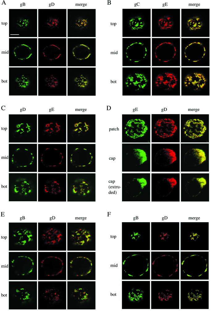
FIG.1.
Copatching of four major PRV glycoproteins (gB, gC, gD, and gE) on the surfaces of PRV-infected SK cells and monocytes. Aggregation of one of these four viral cell surface proteins (patching) by using monospecific antibodies leads to coaggregation of the others (copatching). (A, B, C, and D) SK cells, at 13 h p.i., were incubated with monoclonal antibodies against gB (A), gC (B), gD (C), or gE (D) for 30 min at 37°C and subsequently for 30 min at 37°C with FITC-labeled goat anti-mouse antibodies, leading to patching of gB, gC, gD, or gE. Afterwards, cells were paraformaldehyde fixed and incubated with swine polyclonal anti-gD (A and D) or rabbit polyclonal anti-gE (B and C) antibodies followed by goat anti-swine-Texas red or goat anti-rabbit-Texas red, respectively. The left columns (green) show patched antigen, the middle columns show copatched antigen (red), and the right columns (yellow) show merged image of the former two. In panels A, B, and C, sections through the top, middle (mid), and bottom (bot) of a cell are shown. Panel D shows extended-focus images of cells with patched, capped, and extruded antibody-antigen complexes. (E) Same as panel A but with PRV-infected monocytes instead of SK cells. (F) Same as panel A but without paraformaldehyde fixation. Bar, 5 μm.

