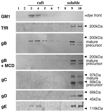
FIG. 4.
A significant amount of PRV gB and a small amount of PRV gE, but no detectable PRV gC or gD, float to lipid raft fractions upon cold Triton X-100 lysis of PRV-infected cells. SK cells, at 13 h p.i., were either first incubated for 45 min at 37°C with 10 mM MCD or immediately lysed with 1% ice-cold Triton X-100. Cell lysates were separated by density ultracentrifugation, and different fractions from top (light) to bottom (heavy) were collected. Samples from each fraction were subjected to SDS-PAGE and Western blotting and analyzed for the presence of GM1, TfR, gB, gC, gD, or gE. Light fractions correspond to lipid raft fractions, and heavy fractions correspond to soluble fractions.

