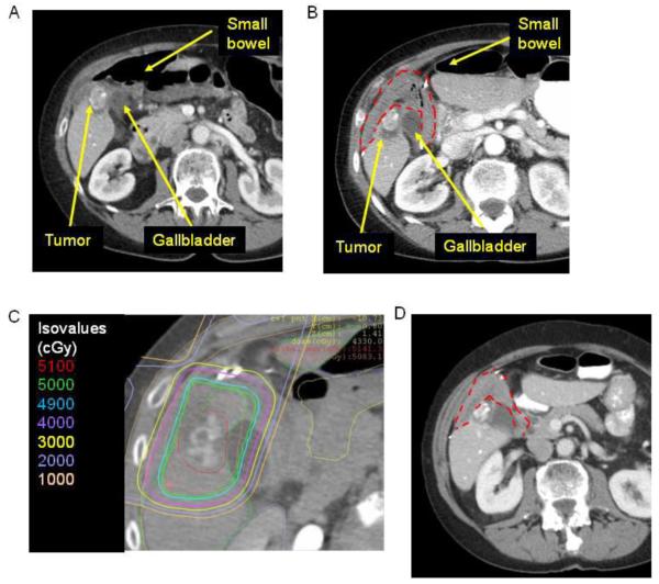
Figure 2.
A) Axial CT image of an intrahepatic cholangiocarcinoma adjacent to the gallbladder and small bowel prior to placement of biological mesh spacer. Tumor is partially calcified. B) Axial CT image following placement of BMS (red dotted line). C) Axial CT image with radiation planning after placement of BMS (red dotted line). D) Axial CT image 18 months after BMS placement and radiation.

