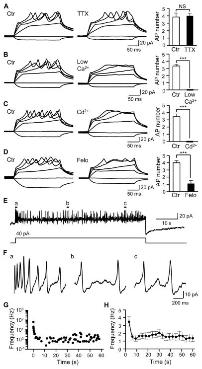
Figure 2. Merkel cells fire slowly adapting Ca2+-action potentials.
(A) Merkel cell APs in the absence (left) and presence (middle) of 0.5 μM TTX. Right panel, summary data (n = 6). (B) Merkel cell APs depend on extracellular Ca2+. Left panel, APs in normal Krebs solution ([Ca2+]o = 2.5 mM). Middle panel, failure to fire APs in a bath solution with low extracellular Ca2+ (20 μM). Right Panel, summary data (n = 6). (C) Merkel cell APs are abolished by Ca2+ channel blocker Cd2+. Left, in the absence of Cd2+; Middle panel, in the presence of 300 μM Cd2+, Right panel, summary data (n = 9). (D) Merkel cell APs are abolished by L-type VGCC blocker felodipine (Felo). Left panel, in the absence of felodipine; Middle panel, in the presence of 0.1 μM felodipine, Right panel, summary data (n = 9). (E) Merkel cell APs in response to a 1-min depolarizing current step at 40 pA. The recording was performed in normal Krebs solution. (F) APs at an expanded scale in the a, b, and c locations indicated in E. (G) Representative plots of instantaneous AP frequency over the 1-min recording shown in E. (H) Summary data for the experiments represented in E. The frequency at each point is calculated with a time bin of 3 sec. Results are pooled from 11 Merkel cells (n = 11). Data represent the mean ± SEM. NS, no significant difference; *** P < 0.001, paired Student t-test. See also Figure S1.

