
Figure 1.
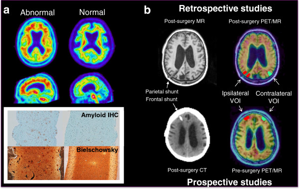
Examples of abnormal and normal [18F]flutemetamol positron emission tomography (PET) and corresponding magnetic resonance (MR) or computed tomography (CT) imaging and histopathology. Panel a) [18F]Flutemetamol PET imaging correlates with histopathology (Study D). Amyloid plaques were determined in biopsy samples by 4G8 imunohistochemistry (IHC). Neuritic plaques were identified in serial sections using a modified Bielschowsky silver stain. Panel b) [18F]Flutemetamol PET images were obtained either retrospectively after biopsy (Studies A and C) or prospectively before biopsy (Studies B and D). Small cortical biopsies were taken during shunt placement and histopathology was correlated to standard uptake value ratio (SUVR) measures in volumes of interest (VOIs) either ipsilateral or contralateral to the site of biopsy.