Figure 1.
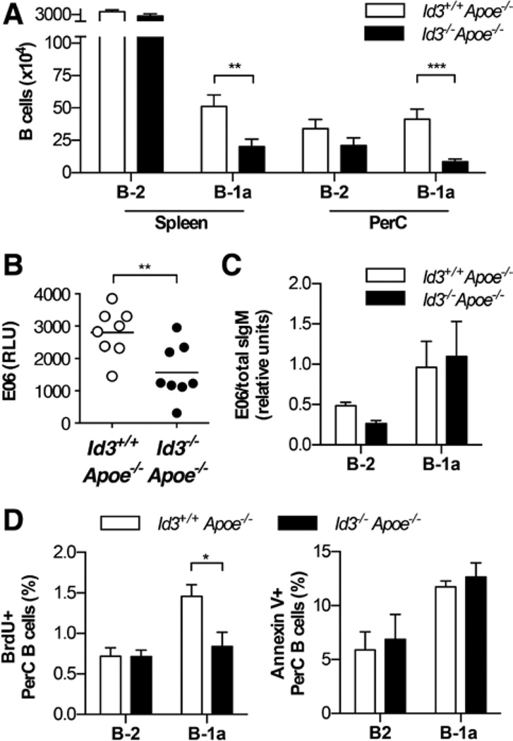
Id3−/−Apoe−/− mice have fewer B-1a B cells in the spleen and peritoneal cavity (PerC) compared with Id3+/+Apoe−/− mice. A, Number of B-2 B cells (CD19+B220hi) and B-1a (CD19+B220loCD5+CD43+IgMhi) in the spleen and PerC of Id3+/+Apoe−/− (n=13) or Id3−/−Apoe−/− (n=12) mice at 8 weeks as measured by flow cytometry. B, E06 levels in the serum of Id3+/+Apoe−/− or Id3−/−Apoe−/− mice as measured by ELISA. C, E06 sIgM transcript levels in peritoneal B cell subsets, B-1a (CD19+B220lo CD5+) and B-2 (CD19+B220hi) B cells, measured by real-time polymerase chain reaction and normalized to total sIgM. D, Bromodeoxyuridine (BrdU) incorporation and annexin V staining in PerC B cell subsets of Id3+/+Apoe−/− (n=6 or 7) or Id3−/−Apoe−/− (n=6 or 7) mice from 3 independent experiments. *P<0.05, **P<0.01, and ***P<0.001.

