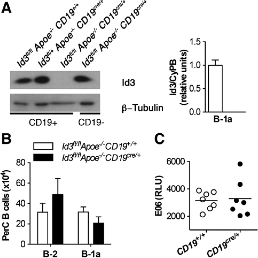
Figure 2.
Generation of a B cell–specific knockout of inhibitor of differentiation 3 (Id3) reveals that the loss of Id3 in B cells does not result in altered numbers of B-1a B cells or E06 levels. A, Left, Id3 and β-tubulin protein in total splenic B cells from Id3fl/flApoe−/−CD19+/+, Id3fl/+Apoe−/−CD19Cre/+, Id3fl/flApoe−/−CD19Cre/+ and Id3fl/fl, Apoe−/−, CD19Cre/+ mice. Right, Peritoneal lavage cells were sorted from Id3fl/flApoe−/−CD19+/+ (open bar) or Id3fl/flApoe−/−CD19Cre/+ (closed bar) mice for B-1a (CD19+B220lo CD5+) cells as in Figure II in the online-only Data Supplement, and Id3 transcript was measured by real-time polymerase chain reaction and normalized to a housekeeping gene, cyclophilin b (CypB). B, Quantification of peritoneal B-2 and B-1a B cell subsets by flow cytometry in Id3fl/flApoe−/−CD19+/+ (n=10) or Id3fl/flApoe−/−CD19cre/+ (n=11) mice at 8 weeks. C, Serum levels of E06 in Id3fl/flApoe−/−CD19+/+ (open circles) or Id3fl/flApoe−/−CD19cre/+ (closed circles) mice were determined by ELISA. Values are the mean relative light units of duplicate determinations.

