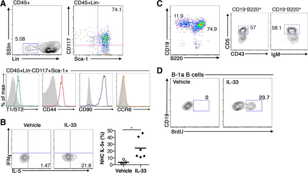
Figure 5.
Natural helper cells are present in the aorta, including periaortic adventitia and perivascular adipose tissue (PVAT), and produce interleukin (IL)-5. Whole aortas containing the adventitia and PVAT were isolated from Apoe−/− mice. A, Representative flow cytometry plots of natural helper (NH) cells (CD45+, lineage negative, CD117+, Sca1+). B, Left, Interferon-γ (IFNγ) and IL-5 intracellular staining in aortic NH cells and right, quantification of the percentage of NH cells expressing IL-5 (n=6 in each group) after vehicle or IL-33 treatment. Representative flow cytometry plots of (C) B cell subsets, B-2 (CD19+B220hi), and B-1a (CD19+B220loCD5+CD43+IgMhi) and (D) BrdU incorporation in aortic B-1a B cells after vehicle or IL-33 treatment. Numbers on flow cytometry plots indicate the percentage of the population of interest. *P<0.05. SSlin indicates side scatter linear.

