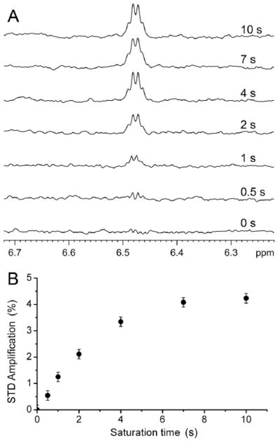
Fig. 3. Saturation transfer difference (STD) spectra of the α7 TM domain for halothane binding.

(A) Prolonged saturation time increased halothane (3.2 mM) signal in the STD spectra in the presence of α7. The STD spectra resulted from the subtraction of the off- (25 ppm; blank region) from the on-resonance (0.4 ppm; protein methyl group) spectra. (B) STD amplification (%) as a function of the saturation time. STD amplification is defined as (Voff − Von)/Voff, Voff and Von are the integrals of halothane peak in the spectra with off- and on-resonance saturation, respectively.
