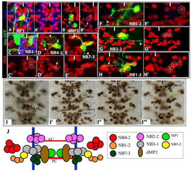
Figure 1. Hb9+ neurons arise from eight different CNS lineages.
(A–H) High magnification ventral views of CNS lineage clones (green) that contain Hb9+ neurons (red); FasII (blue) labels axon fascicles allowing identification of the axon tracts followed by GFP-labeled axons. Arrowheads identify the motor neuron exit(s). (A) Two-cell clone of sibling Hb9+ MP1 intersegmental interneurons. (B) Two-cell clone of d/vMP2 sibling neurons; dMP2 visceral motor neuron (arrow) expresses Hb9. (C) A partial clone of the NB 3-1 lineage contains three Hb9+ motor neurons. (D) A lineage clone of NB4-2 contains the well-characterized Hb9− RP2 motor neuron (asterisk) and three Hb9+CoR motor neurons (arrow); two Hb9+interneurons located ventral to the CoRs are not visible at this focal plane. (E) The well-characterized NB 7-3 lineage generates three EW interneurons, which express Hb9, and the Hb9−GW motor neuron. (F) A single-cell clone of the motor neuron in theNB5-2 lineage extends an axon contralaterally out of the CNS. (G) NB2-2 generates a large clone of motor neurons and interneurons; 6 ventral Hb9+ interneurons reside in the NB2-2 lineage. (H) Lineage clone of NB1-2 contains only interneurons, four of which express Hb9 and project axons contralaterally via the posterior commissure (arrow). (I) Ventral to dorsal views of Hb9+ neurons in abdominal segments with the lineal identity of each cluster shown. (J) Schematic representation of Hb9+cell lineages. White or black line mark the midline of the CNS.

