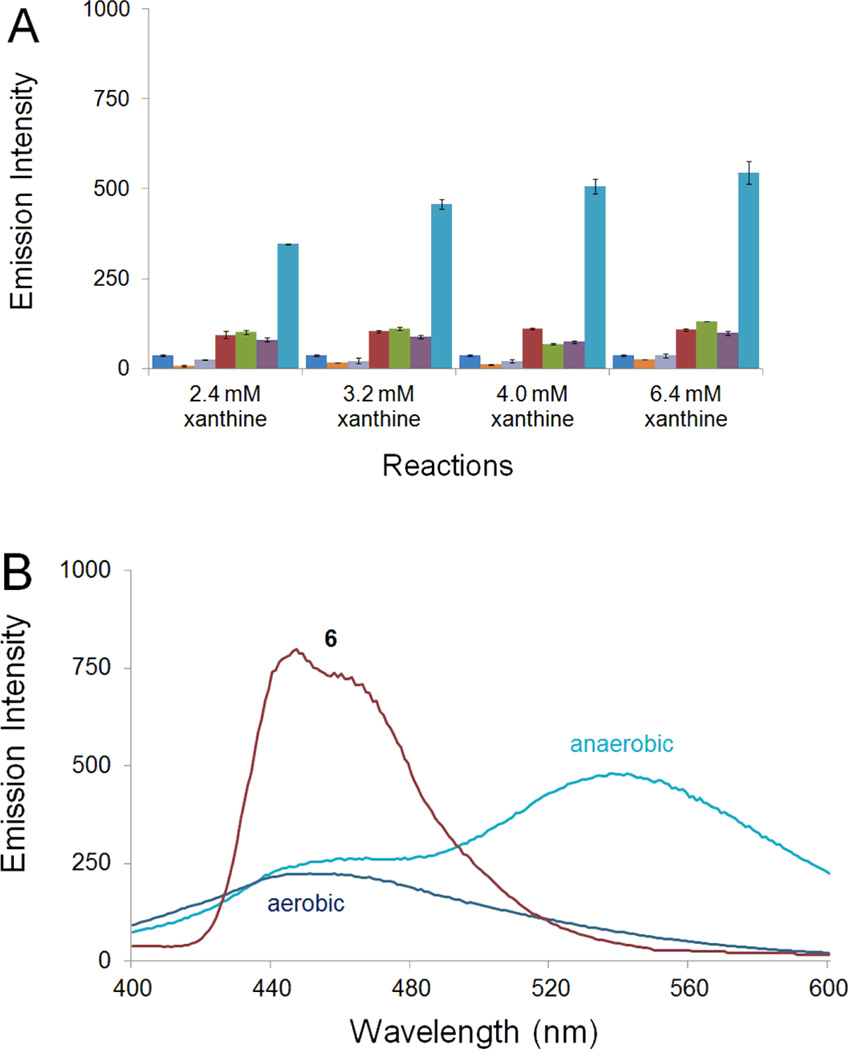
Figure 6.
Enzymatic conversion of 6 into a fluorescent product selectively under hypoxic conditions. A. Fluorescence emission at 530 nm (λex 340 nm). Each set of assays depicted in the bar graph consists of (from left to right): a control sample of compound 6 alone (0.8 mM)  , a control reaction composed of xanthine oxidase (2.4 U/mL) and xanthine (2.4 mM, 3.2 mM, 4.0 mM and 6.4 mM) under aerobic
  , a control reaction composed of xanthine oxidase (2.4 U/mL) and xanthine (2.4 mM, 3.2 mM, 4.0 mM and 6.4 mM) under anaerobic conditions
  , a control reaction composed of xanthine oxidase (2.4 U/mL), xanthine (2.4 mM, 3.2 mM, 4.0 mM and 6.4 mM) and the non-fluorescent electron acceptor, 1,2,4-benzotriazine 1,4-dioxide, (6.4 mM) under aerobic conditions
  , a control reaction composed of xanthine oxidase (2.4 U/mL), xanthine (2.4 mM, 3.2 mM, 4.0 mM and 6.4 mM) and the non-fluorescent electron acceptor, 1,2,4-benzotriazine 1,4-dioxide, (6.4 mM) under anaerobic conditions
  , a reaction composed of xanthine oxidase (2.4 U/mL), xanthine (2.4 mM, 3.2 mM, 4.0 mM and 6.4 mM) and 6 under aerobic conditions
  , a reaction composed of xanthine oxidase (2.4 U/mL), xanthine (2.4 mM, 3.2 mM, 4.0 mM and 6.4 mM) and 6 under anaerobic conditions
  . Reactions were incubated for 18 h in sodium phosphate buffer at (12 mM, pH 7.4) at 24 °C, diluted with aerobic sodium phosphate buffer (12 mM, pH 7.4), and the fluorescence measured (λex 340 nm, λem 530 nm). B. Fluorescence spectra of aerobic and anaerobic reaction mixtures containing 6 (0.8 mM), xanthine oxidase (2.4 U/mL), and xanthine (3.2 mM) and fluorescence spectrum of 6 alone (0.05 mM, λex 340 nm, in sodium phosphate buffer, 10 mM, pH 7.4).

