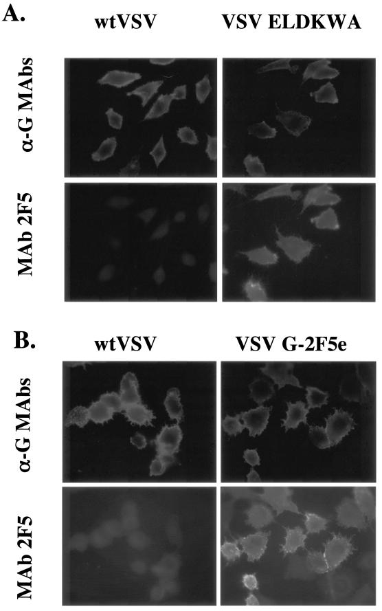
FIG. 7.
Indirect immunofluorescence microscopy of cells infected with VSV, VSV 191 G-ELDKWA, or VSV G-2F5e. BHK cells were infected with wt VSV or VSV 191 G-ELDKWA (A), or cells were infected with wt VSV or VSV G-2F5e (B). Cells were fixed and then incubated with mouse MAbs I1 and I14 to VSV G (α-G MAbs) and with the human MAb 2F5 as indicated. Cells were then incubated with a Texas red-conjugated anti-mouse antibody and a FITC-conjugated anti-human antibody. Cells were photographed as described in the legend to Fig. 4.

