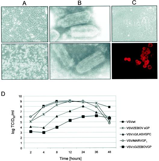
FIG. 2.
Characterization of rescued rVSVs. Rescued rVSVs were used to infect VeroE6 cells at an MOI of 0.1 PFU/cell. (A) Cytopathogenic effect of infected VeroE6 cells is shown by phase-contrast microscopy 24 h postinfection with VSVΔG/MARVGP (lower panel) in comparison with a mock-infected culture (upper panels). (B) Particle morphology. Electron micrographs show wild-type VSV (upper panel) and VSVΔG/MARVGP (lower panel). (C) Immunofluorescence staining of VeroE6 cells infected with VSVΔG/MARVGP with the GP-specific monoclonal antibody 5EII (dilution, 1:1,000) (lower panel). The upper panel shows the same cells in bright-field microscopy. (D) Growth curves. VeroE6 cells were infected with wild-type VSV (VSVwt), VSV/ZEBOVsGP, VSV/MARVGP1, VSVΔG/ZEBOVGP, VSVΔG/LASVGP, or VSVΔG/MARVGP at an MOI of 10. Supernatants were collected at the indicated times and titrated by defining the TCID50.

