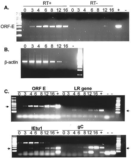
FIG. 2.
Detection of ORF-E RNA by RT-PCR. MDBK cells were infected with the Cooper isolate of BHV-1 (multiplicity of infection of 5). Total RNA was extracted at the indicated times postinfection. (A) Strand-specific RT-PCR was conducted with primer 308 (Fig. 1C) to synthesize cDNA. The primers for PCR were 308 and 598. (B) With the same RNA as shown in panel A, cDNA synthesis was primed with random primers, and β-actin cDNA was amplified with the primers described in Materials and Methods. Reactions without reverse transcriptase did not yield specific amplified products with the β-actin primers (data not shown). (C) Random primers were used to synthesize cDNA. PCR was then performed with specific primers for ORF-E (308 and 598), the LR gene (L3B), IETU1, and gC. Lanes +, positive controls consisting of DNA from infected cells; lanes −, no-template controls; RT−, no reverse transcriptase enzyme in the reaction. PCR products were separated on a 2% agarose gel, and the products were visualized with ethidium bromide. The numbers above the lanes indicate hours after infection. The arrows denote products of the expected sizes from the PCR.

