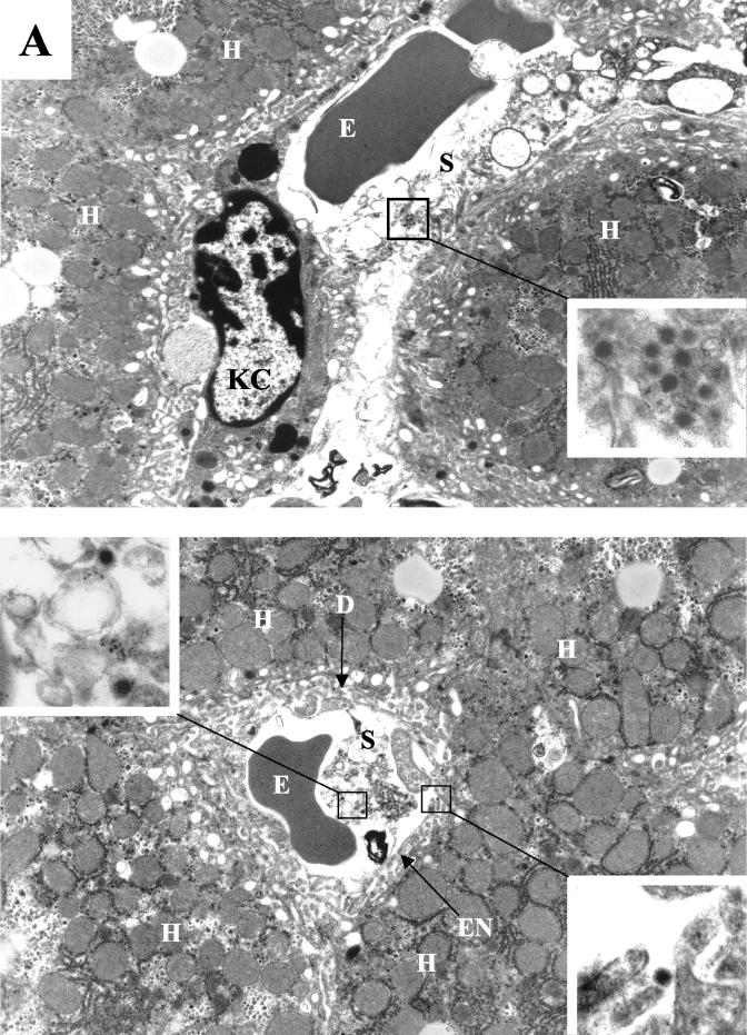
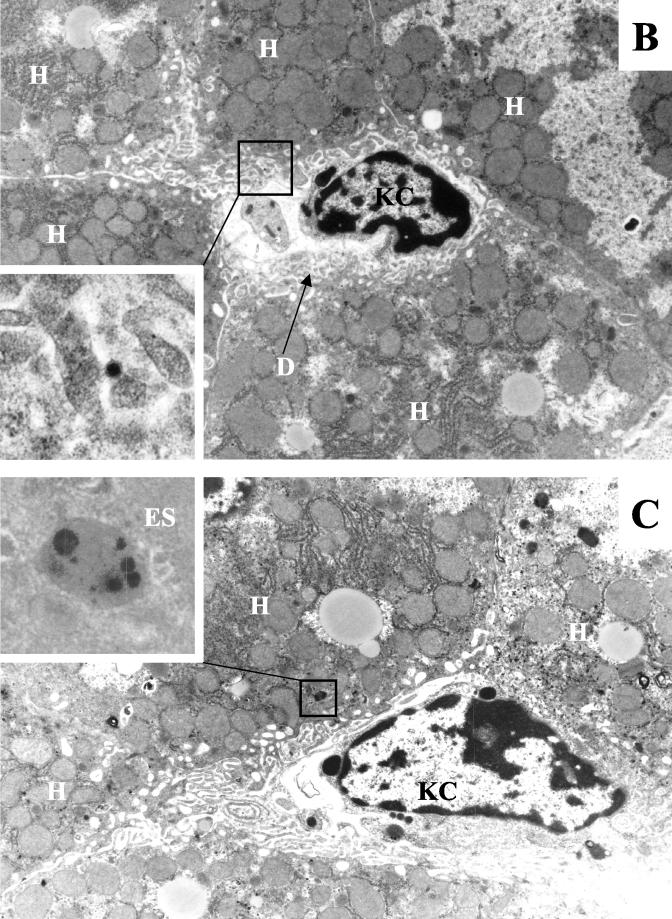
FIG. 3.
Visualization of Ad distribution in liver tissue 30 min after intravenous application. (A) Ads present in hepatic sinusoids. (Upper panel) Ad5/9L. (Lower panel) Ad5/9S. (B and C) Ad particles present in the space of Disse (B) and in hepatic endosomes (C) in liver sections from Ad5/9L-injected mice. Note that Ad5/9S particles were present both in sinusoids and in the space of Disse (A, lower panel, upper and lower insets, respectively). H, hepatocyte; KC, Kupffer cell; S, sinusoid; E, erythrocyte; D, space of Disse; EN, sinusoid endothelial cell; ES, endosome. The characteristic morphology of Ad particles can be seen clearly on the enlarged images of selected areas. Magnifications: ×4,400 for main images and ×21,000 for enlarged images.


