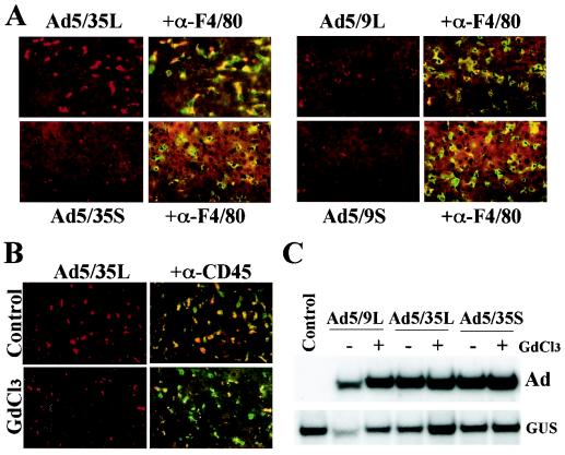
FIG. 6.
Association of Ad vectors with Kupffer cells (A and B) and liver cell transduction in Kupffer cell-depleted mice (C). (A) Cy-3-labeled Ads were injected into the tail vein of C57BL/6 mice. At 30 min later, livers were recovered and immediately frozen in OCT compound. To visualize Kupffer cells, fixed liver sections were stained with anti-F4/80 antibody (green). Cy-3-labeled virus appears red. Images of representative fields were taken with red and green filters and then were superimposed to reveal the association of Ad vectors with Kupffer cells (yellow). (B) Mice were treated with gadolinium chloride (GdCl3) to functionally inactivate Kupffer cells, which were stained with anti-CD45 antibody (green). At 30 min after injection of Cy-3-labeled Ads, livers were recovered, and the association of Ad vectors with Kupffer cells was evaluated. Note that in GdCl3-treated mice, the association of Ad vectors with Kupffer cells was dramatically reduced. (C) Southern blot analysis of Ad genomes present in the livers of mice treated with GdCl3 and normal mice (−GdCl3) 30 min after systemic vector application. GUS, mouse β-glucuronidase gene. The control was liver DNA from a mouse injected with virus dilution buffer (PBS) only.

