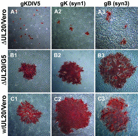
FIG. 3.
Plaque morphology of UL20-null viruses on Vero cells (A), UL20-null viruses on G5 cells (B), and UL20-null rescued viruses on Vero cells (C). Confluent cell monolayers were infected with Δ20gKDIV5 virus (A1 and B1), Δ20syn20DIV5 virus (A2 and B2), or Δ20gBsyn3 virus (A3 and B3) or the corresponding UL20-null rescued virus (C1, C2, and C3) at an MOI of 0.001, and viral plaques were visualized by immunohistochemistry at 36 hpi.

