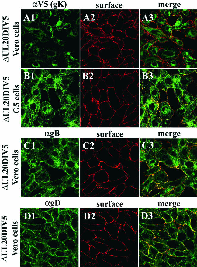
FIG. 4.
Confocal microscopic visualization of cell surface and intracellular distribution of gK (A and B), gB (C), and gD (D) in either Vero (A, C, and D) or G5 (B) cells infected with UL20-null Δ20gK DIV5 at an MOI of 10. Infected cell surfaces were labeled with biotin (red) under live conditions. Cells were fixed and processed for confocal microscopy and labeled with anti-gD (αgD, green) (D1 and D3), anti-gB (αgB, green) (C1 and C3), or anti-V5 (αV5) for gK (green) (A1, A3, B1, and B3). Superimpositions of red and green images for each group are shown (A3, B3, C3, and D3).

