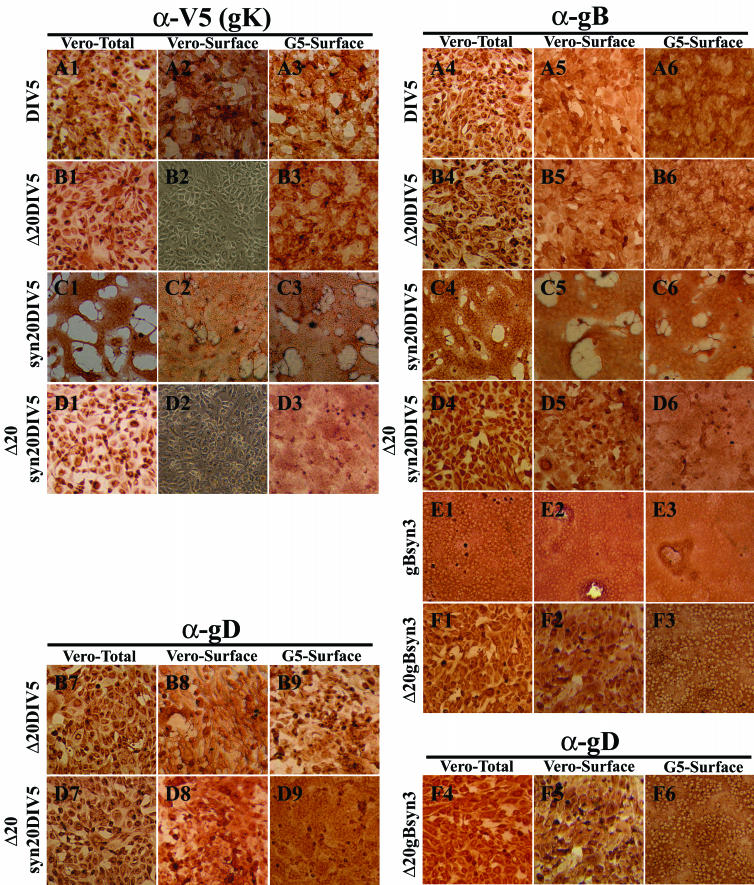
FIG. 5.
Cell surface immunohistochemical detection of gK (A1 to A3, B1 to B3, C1 to C3, and D1 to D3), gB (A4 to A6, B4 to B6, C4 to C6, D4 to D6, E1 to E3, and F1 to F3), or gD (B7 to B9, D7 to D9, and F4 to F6). Vero (1, 2, 4, 5, 7, and 8) or G5 (3, 6, and 9) cells were infected with DIV5 (A1 to A6), Δ20DIV5 (B1 to B9), syn20DIV5 (C1 to C6), Δ20syn20DIV5 (D1 to D9), gBsyn3 (E1 to E3), or Δ20gBsyn3 (F1 to F6) at an MOI of 10, and cells were immunohistochemically processed at 12 hpi under either live (2, 3, 5, 6, 8, and 9) or fixed and permeabilized (1, 4, and 7) conditions. α-V5, anti-V5; α-gB, anti-gB; α-gD, anti-gD.

