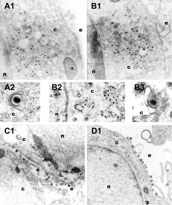
FIG. 6.
Electron micrographs of Vero (A and B) and G5 (C and D) cells infected with either Δ20DIV5 (A and C) or Δ20D3prtC (B and D) virus. Confluent cell monolayers were infected at an MOI of 5, incubated at 37°C for 16 h, and prepared for transmission EM. Nuclear (n), cytoplasmic (c), and extracellular (e) spaces are marked.

