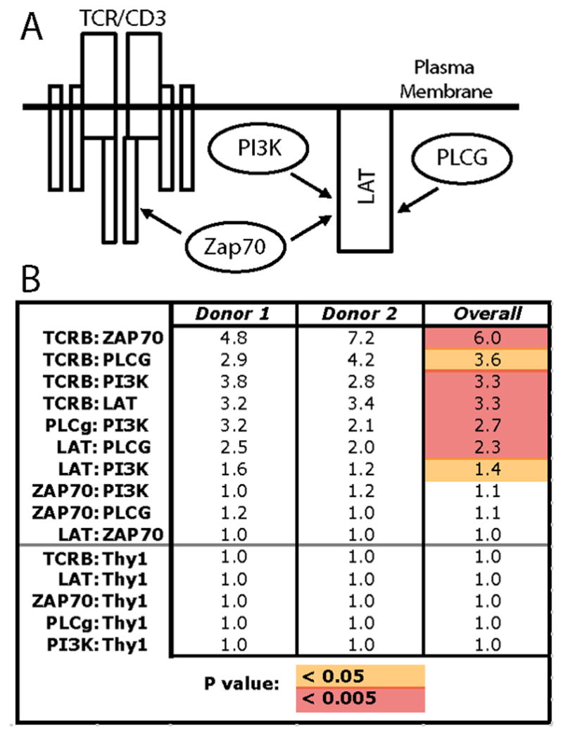
Figure 2. Protein complex analysis from primary human sr-T cells using mIP-FCM.

A) Target proteins are known to form shared complexes that mediate signal transduction upon T cell antigenic stimulation. B) Skin-resident T cells from 2 separate individuals were isolated via the crawl-out method, and then either left unstimulated, or stimulated with anti-CD3 + anti-CD28 for 5 minutes. Following lysis, mIP-FCM was was performed. Numbers represent the fold-change of protein pairs observed in shared complexes in stimulated vs unstimulated conditions. For unified analysis of data from both donors, their corresponding fold-changes were averaged, and raw values of stimulated vs. unstimulated conditions were compared by Student’s t-test (Overall; orange, p < 0.05; red, p < 0.005).
