
Fig. 2.
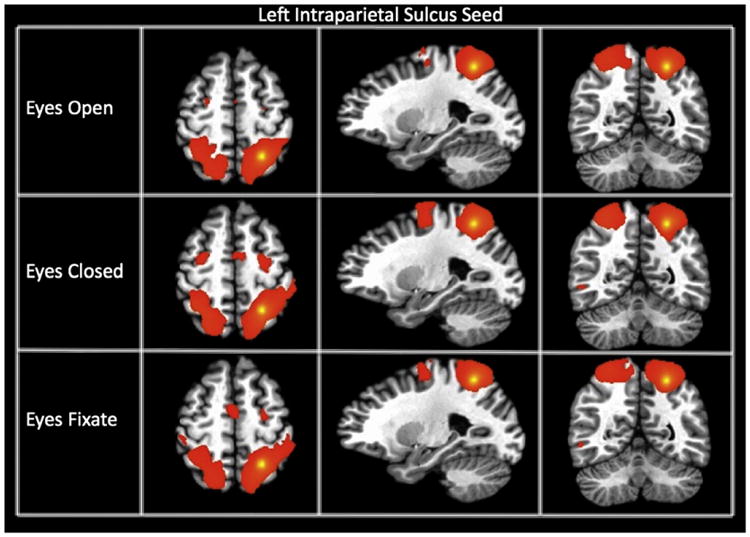
Connectivity maps of the left intraparietal sulcus seed (showing the dorsal attention network) for each resting state condition. The maps are thresholded at a p-value of 1 * 10−15.
Official websites use .gov
A
.gov website belongs to an official
government organization in the United States.
Secure .gov websites use HTTPS
A lock (
) or https:// means you've safely
connected to the .gov website. Share sensitive
information only on official, secure websites.
Connectivity maps of the left intraparietal sulcus seed (showing the dorsal attention network) for each resting state condition. The maps are thresholded at a p-value of 1 * 10−15.