
Figure 1.
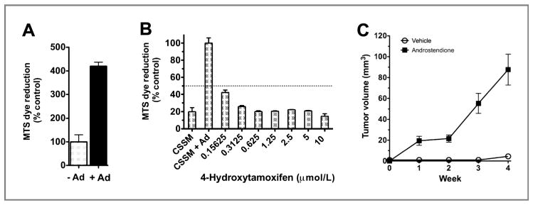
MCF-7/AC-1 cells are estrogen-driven in vitro and estrogen-dependent in vivo. Cells were cultured in IMEM steroid-reduced medium without phenol red for 2 days before plating. Cell proliferation was measured using an MTS assay, as described in Methods. A, effect of presence of androstenedione (Ad) on MCF-7/AC1 cell growth. Cell growth is expressed as the percentage of the cells compared with the control wells (untreated cells). Columns, mean; bars, SE. B, antiproliferative effect of increasing concentrations of 4-hydroxytamoxifen in the presence of 1 nmol/L androstenedione on MCF-7/AC1 cells. Cell proliferation is expressed as the percentage of the cells compared with the control wells (1 nmol/L androstenedione-treated cells). CSSM, untreated cells cultured in steroid-reduced medium. Columns, mean; bars, SE. C, MCF-7/AC-1 xenografts, each mouse received subcutaneous inoculations in 2 sites per flank with 100 μL of MCF-7/AC-1 cell suspension containing 2.5 × 10⁶ cells. The mice were injected daily with supplemental androstenedione (100 μg/d) or vehicle from day 0. Tumors were measured with calipers weekly throughout experiment. Points, mean; bars, SE.