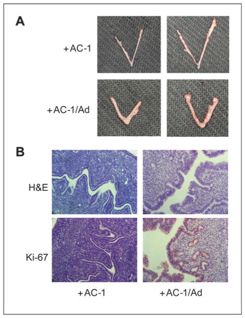
Figure 2.
Uterotropic effects of mice bearing MCF-7/AC-1 tumors, with and without androstenedione. Each mouse received subcutaneous inoculations in 2 sites per flank with 100 μL of MCF-7/AC-1 cell suspension containing 2.5 × 10⁶ cells. The mice were injected daily with supplemental androstenedione (100 μg/d) or vehicle from day 0. At day 28, the mice were sacrificed and uteri were removed from the mice, weighed, formalin-fixed, and embedded in paraffin. A, morphologic appearances of 2 representative uteri from ovariectomized MCF-7/AC-1 xenografts bearing mice were shown with or without androstenedione for 28 days. B, analysis of cell proliferation. Uterine cross-sections were examined by Ki-67 immunostaining. Reddish nuclear deposits indicate the sites of positive immunostaining (20×).

