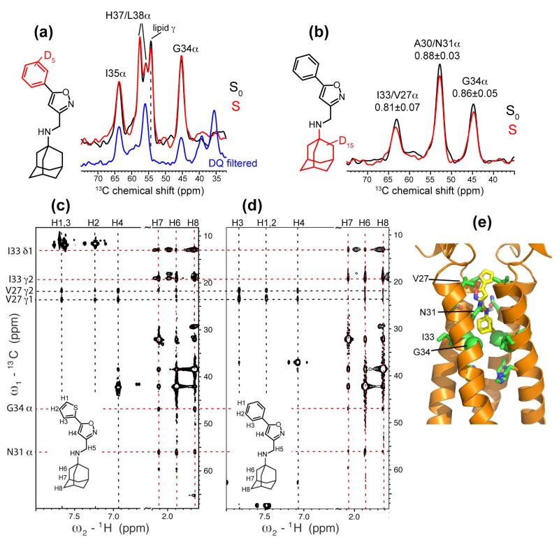
Figure 6.
Binding site and orientation of WJ352 in S31N bound to DMPC bilayers (a, b) and DPC micelles (c, d). (a) 13C-2H REDOR S0 (black) and S (red) spectra of d5-WJ352 complexed GIHL22-46, S31N in DMPC bilayers. The mixing time was 14.2 ms and the drug : tetramer ratio was 8 : 1. The deuterated phenyl group did not dephase any peptide 13C signals, especially G34. The REDOR spectra were measured without DQ filtering, thus showed a lipid headgroup Cγ peak at 54 ppm. This is verified by the absence of the peak in a 13C DQ filtered spectrum (blue). (b) 13C-2H DQ filtered REDOR spectra of d15-WJ352 bound VANIG19-49, S31N in DMPC bilayers. The mixing time was 9.6 ms and the drug : tetramer ratio was 1 : 1. The deuterated adamantane dephased multiple 13C signals. (c, d) 2D 13C-(1H)-1H NOESY (150 ms) spectra of VANIG19-49, S31N in DPC micelles with bound WJ332 (c) or with bound WJ352 (d). Drug – peptide cross peaks are observed and consistent with the amine-up orientation. (e) Solution NMR structure of S31N-M2 with bound WJ332 (PDB: 2LY0)29.

