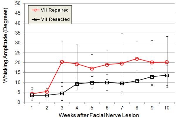
Figure 3.
Average whisking amplitude is shown for the 3 largest whisks across weeks 1-10 of recovery, ipsilateral to facial nerve transection and suture repair (VII Repaired group, open circles in red; N=9) versus ipsilateral to facial nerve resection (VII Resected group, open boxes in black, N=7). Group averages were statistically compared for weeks 3, 4, 6, 7, and 10, and significantly differed at weeks 3, 4, and 6, but not at weeks 7 and 10. Error bars are ± 1 standard deviation.

