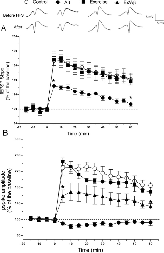
Figure 2.
Hippocampal early phase LTP (E-LTP) measured as increases in the slope of the fEPSP (A) and pspike amplitude (B) in area CA1 was evoked by HFS (applied at time zero) of the Schaffer collateral synapses in anesthetized rats. In rats with Aβ1–42 infusion (Aβ), E- LTP was significantly more impaired than other groups. Ex/Aβ rats exhibited a similar fEPSP slope compared to that of control and exercised rats. Additionally, the pspike amplitude of Ex/Aβ rats was markedly different from those of Aβ rats. Each point is the mean ± SEM of 5–6 rats. Points between the two asterisks (*) indicate significant difference from all groups.

