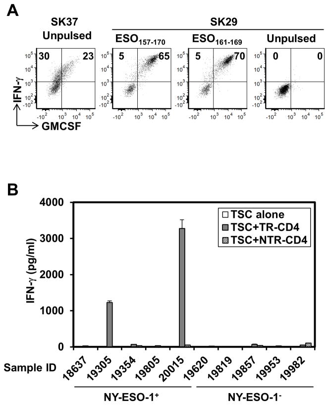
Figure 3.
Detection of TR-CD4 at the local tumor site. (A) Tumor-infiltrating lymphocytes from an ovarian cancer patient were stimulated with NY-ESO-1157–170. After 20-days, NY-ESO-1157–170-reactive CD4+ T cells were isolated and expanded. Recognition of SK37, NY-ESO-1157–170, and NY-ESO-1161–169 was tested by intracellular staining. Values in quadrants indicate percentages of cells. (B) TR-CD4 or NTR-CD4 cells were co-cultured with NY-ESO-1+/− tumor single cell suspensions (TSC) obtained from DP04+ patients for 24 hours. IFN-γ level in the supernatant was measured by ELISA.

