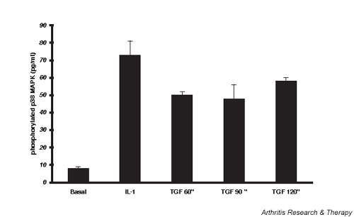
Figure 1.

Lapine chondrocyte p38 mitogen-activated protein kinase (MAPK) phosphorylation. Chondrocytes were grown to confluence, medium serum reduced for 24 hours, and the cells lysed 60–120 min after activation with transforming growth factor beta (TGF-β) or 30 min after activation with IL-1. ELISA for phospho-p38 MAPK in the lysates was done as per instructions in the R&D Systems kit. Data were normalized to the average protein content of the lysates of 33 ± 1 μg/ml. Values are plotted as mean pg/ml ± standard error of n = 3–10.
