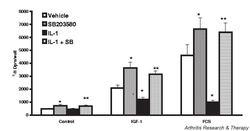
Figure 4.

Inhibition of p38 mitogen-activated protein kinase potentiates basal and growth factor-stimulated proliferation of rabbit chondrocytes, and reverses IL-1-inhibited proliferation. Chondrocytes were grown to 80% confluence, the medium serum reduced for 24 hours, SB 203580 (SB) (1 μM) or dimethyl sulfoxide vehicle (0.5%) added, and 2 ng/ml IL-1 added 30 min later. The cells were then stimulated with insulin-like growth factor 1 (IGF-1) (50 ng/ml) or fetal calf serum (FCS) (10%) for 24 hours. Proliferation was assayed as [3H]thymidine incorporation into trichloroacetic acid precipitated material following a 2 hour pulse label. Values are the mean ± standard error of n = 10–20. *P < 0.05 versus vehicle, **P < 0.05 versus IL-1.
