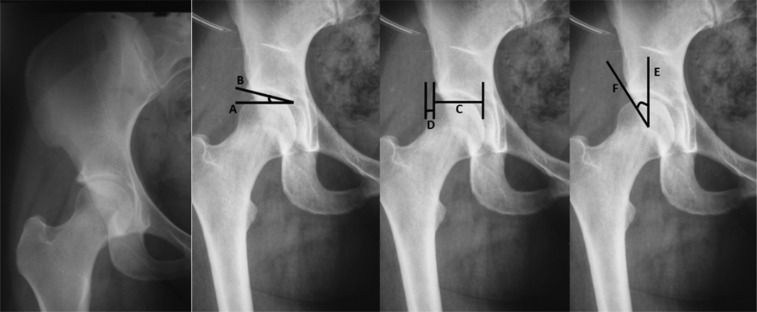
Figure 3.
A) Anteroposterior X‐ray of a normal hip; B) Acetabular index–angle formed by a horizontal line (line A) and a line connecting the medial point of the sclerotic zone with the lateral edge of the acetabulum (line B); C) Femoral head extrusion index–line C corresponding to the connection of both extremities of the acetabulum esclerotic zone and line D corresponding to the connection of the femoral head‐neck junction and the sclerotic zone of the acetabulum; and D) Lateral center edge angle of Wiberg–a vertical line from the femoral head center (line E) and a line connecting the femoral head center with the lateral edge of the acetabulum (line F).

