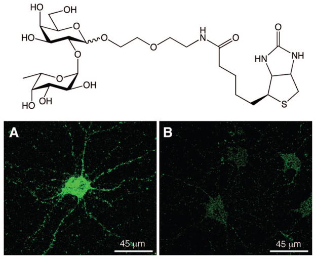
Figure 8.
Chemical probe for imaging lectin receptors (top) and staining of hippocampal neurons in culture (bottom panels) with the probe demonstrating the presence of Fucα(1–2)Gal lectins along the cell body and neurites. Cells were treated with 3 mM of the imaging probe (A) or biotin (B), labeled with a streptavidin–dye conjugate, and imaged by fluorescence microscopy. Images courtesy of C. Gama.

