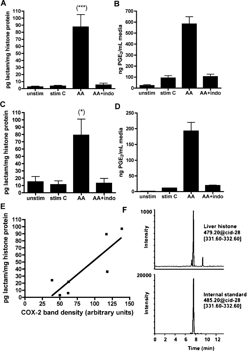
Figure 2.
LG-lysine adducts are found in cells and tissue, dependent on COX-2 activity. RAW264.7 mouse macrophage (A) and A549 human lung carcinoma (C) cells were stimulated to express COX-2 and then given 20 μM arachidonic acid (AA) or vehicle. A subgroup of cells was preincubated 45 min with 50 μM indomethacin. As a measure of COX activity, PGE2 was determined by GC–MS from cell media prior to lysis (B and D). Nuclei were isolated, and histones were extracted and digested to individual amino acids prior to LC–ESI/MS/MS analysis. *p < 0.05; ***p < 0.001 by ANOVA followed by Tukey’s post-test (n ≥ 5). (E). Histones were extracted from nuclei of rat liver and analyzed as above for LG-lactam adduct. COX-2 protein was analyzed by Western blotting and plotted against lactam adduct levels. Each point corresponds to one liver, and shown is the line of regression (r2 = 0.7237). Pearson r = 0.8507; two-tailed p = 0.0152. (F) LC–MS chromatograph of histones isolated from a rat liver with relatively high COX-2 expression (COX-2 band intensity of 117 arbitrary units).

