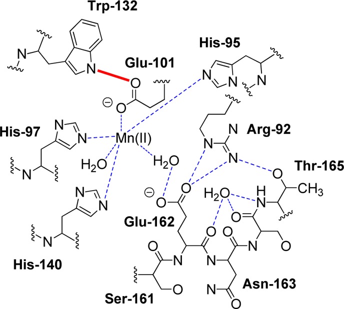
Figure 3.

Schematic representation of hydrogen bonding interactions (dotted lines) involving residues in the Bacillus subtilis OxDC N-terminal Mn(II) binding site when the active site loop adopts a “closed” conformation.10a The Glu-101/Trp-132 hydrogen bond is shown in red. Reproduced with permission from ref (10a). Copyright 2004 American Society for Biochemistry and Molecular Biology.
