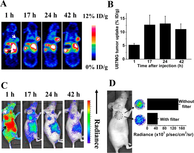
Figure 3.

(A) Representative whole-body coronal PET images of U87MG tumor-bearing mice at 1, 17, 24, and 42 h after intravenous injection of 250 μCi of 64Cu-doped QD580 (n = 3). White arrow, tumor area; black arrow, liver area. Slices for the images are 1 mm thick. QDs show typical reticuloendoethlial system uptake in the liver and spleen as well as tumor accumulation via an EPR effect. (B) ROI analysis of U87MG tumor uptake of 64Cu-doped QDs over time (n = 3). (C) Representative whole-body luminescence images of U87MG tumor-bearing mice at 1, 17, 24, and 42 h postinjection of 250 μCi of 64Cu-doped QD580 (n = 3). White arrow, tumor area; black arrow, liver area. (D) Comparison of photon flux obtained via an open window without filter and that obtained via a filter covered from 575 to 650 nm in the tumor area 42 h postinjection of 250 μCi of 64Cu-doped QDs.
