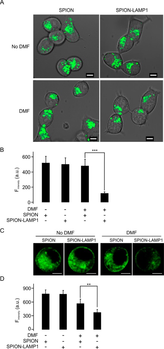
Figure 4.

DMF treatment decreases intracellular lysosomes and the pH in LAMP1-SPION loaded INS-1 cells. (A) The cells were treated by DMF (20 Hz, 20 min) and lysosomes stained with LysoTracker Green. Scale bars = 5 μm. (B) Mean intensity of fluorescence was measured under the different conditions in (A). (C) Representative confocal images indicate the intracellular pH value using an acidotropic probe, LysoSensor Green DND 189 in INS-1 cells. Scale bars = 5 μm. (D) Mean fluorescence intensities were measured as in (C). Data collected from five experiments with at least 6 cells under each condition. **p < 0.01, ***p < 0.001.
