
Figure 4.
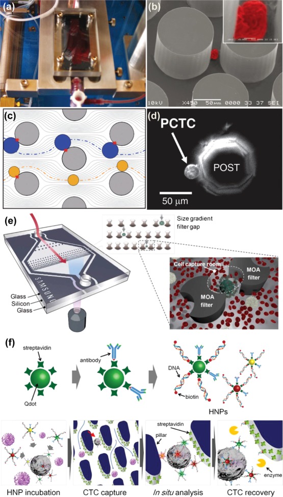
Silicon-based microfluidic CTC separation technologies. (a and b) Setup of post-based CTC-chip developed by Nagrath et al. Captured NCI-H1650 cell imaged with scanning electron microscopy (color added) with high magnification inset.46 Adapted with permission from ref (46). Copyright 2007 Nature Publishing Group. (c and d) Computer simulated particle paths for larger cells (blue) compared with smaller cells (yellow) for geometrically enhanced differential immunocapture (GEDI). Prostate cancer tumor cell captured on octagonal post of GEDI microdevice.49 Adapted with permission from ref (49). Copyright 2010 The Royal Society of Chemistry. (e) Filtration unit with two different filter gaps for the capture of CTCs that have undergone size amplification with magnetic particles.52 Adapted with permission from ref (52). Copyright 2012 The Royal Society of Chemistry. (f) Hybrid nanoparticles consisting of antibodies, quantum dots, and specific DNA sequences for the labeling, capture, and release of CTCs in a capture and recovery chip.53 Adapted with permission from ref (53). Copyright 2013 Elsevier.