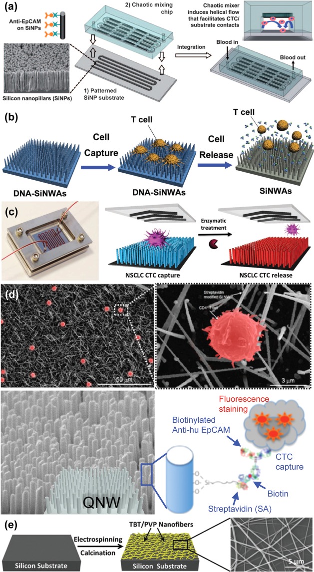
Figure 9.
Nanopillar, nanowire, and nanofiber structures. (a) Chaotic micromixer induces increased contact between flowing cells and anti-EpCAM functionalized silicon nanopillars (SiNPs) substrates.91 Adapted with permission from ref (91). Copyright 2011 John Wiley & Sons, Inc. (b) T lymphocyte cell capture on DNA-silicon nanowire arrays (SiNWAs) and cell release using exonuclease I to break down aptamers.93 Adapted with permission from ref (93). Copyright 2011 John Wiley & Sons, Inc. (c) Aptamer-coated NanoVelcro Chip for capturing and releasing NSCLC CTCs94 Adapted with permission from ref (94). Copyright 2013 John Wiley & Sons, Inc. (d) CD4+ T lymphocytes (shown by SEM images) may be selectively captured by quartz nanowires (QNWs) functionalized with anti-EpCAM.95,96 Adapted with permission from refs (95) and (96). Copyright 2010, 2012, American Chemical Society. (e) Titanium nanofibers are fabricated through electrospinning and calcination prior to functionalization for ultimate use in cell capture.98 Adapted with permission from ref (98). Copyright 2012 John Wiley & Sons, Inc.

