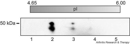
Figure 1.

Purification of placental Sa antigen. The antigen was purified by anion-exchange chromatography from extracts of placenta and subsequently purified by two-dimensional gel electrophoresis according to a three-step procedure described by Liang and colleagues [20]. First, proteins were separated by molecular weight, then proteins of appropriate molecular weight were separated by isoelectric focusing (IEF), and finally proteins with appropriate pI were separated once more by molecular weight. Each step of the procedure was monitored by western blotting with an anti-Sa reference serum. Shown here is the final gel, which was stained with Coomassie brilliant blue. The double band in lane 2 is the Sa antigen that was cut out and used for microsequencing. Each lane represents a portion of the IEF gel (approximate pI is listed above each lane).
