
Figure 5. Anti-TLR4 treatment decreases Cox-2 protein expression and TXB2 release in mesenteric arteries.
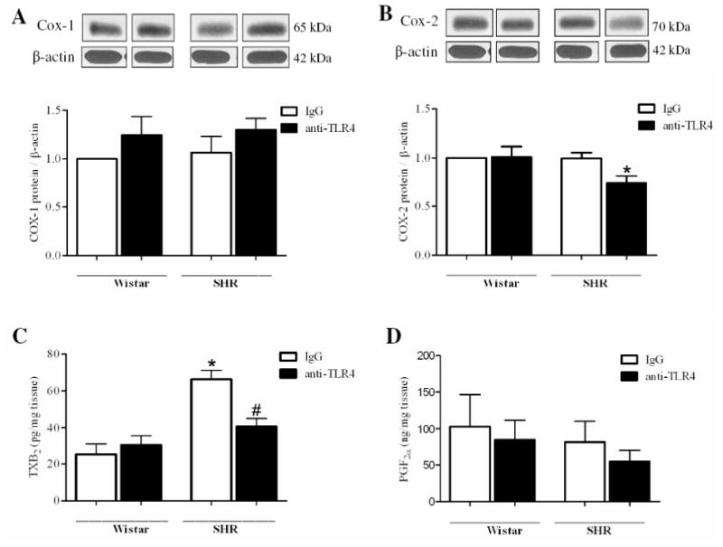
(A) COX-1 and (B) COX-2 proteins expression in mesenteric resistance arteries from IgG-(white bars) or anti-TLR4-treated (black bars) Wistar and SHR rats. On top, representative western blot images of Cox-1 (A) and Cox-2 (B) protein expression. Bar graphs show the relative expression of Cox-1 and Cox-2 after normalization to β-actin expression. Release of (C) thromboxane B2 and (D) 6-keto-prostaglandin F1α by mesenteric arteries stimulated with noradrenaline 100 μM. Each bar represents the mean ± SEM, n = 5-6. *P< 0.05. Statistical test: one-way ANOVA.